Checking Out the Vital Services Offered by a Veterinary Cardiologist: Recognizing Ultrasound and CT Scan Strategies
Veterinary cardiologists play an important duty in the health of pets by detecting and treating various heart conditions. They utilize advanced imaging techniques, such as cardiac ultrasound and CT scans, to provide accurate evaluations. Each approach has its distinctive benefits and applications. Recognizing these methods is necessary for family pet proprietors seeking the most effective take care of their friends. What aspects should family pet owners consider when picking in between these diagnostic tools?

The Function of Vet Cardiologists in Pet Health Care
Vet cardiologists play an important function in the medical care of pets, focusing particularly on detecting and treating heart-related conditions. They have specialized training that permits them to interpret complicated analysis examinations and identify various cardio concerns. These professionals utilize sophisticated strategies, such as echocardiography and electrocardiography, to assess heart function and structure accurately.Veterinary cardiologists additionally create tailored treatment plans that may consist of drugs, way of living modifications, and, sometimes, surgical treatments. Their proficiency prolongs to enlightening pet dog owners about heart health, highlighting the value of normal exams and very early discovery of potential problems. Collaboration with general veterinarians is crucial, as it ensures thorough take care of animals with thought cardiac problems. By providing specialized services, vet cardiologists substantially enhance the high quality of life for family pets and offer comfort for their owners, reinforcing the relevance of heart wellness in total pet dog health.
Typical Cardiac Concerns in Pet Dogs
Usual heart concerns in pet dogs can substantially influence their wellness and high quality of life. Heart murmurs, various sorts of cardiomyopathy, and hereditary heart problems are among the most widespread problems that veterinarians encounter. CT Scans For Animals. Comprehending these issues is important for pet proprietors to assure prompt diagnosis and ideal treatment
Heart Murmurs in Pets
Heart whisperings can be a resource of worry for pet owners, they are not constantly a measure of major wellness issues. A heart whispering is an abnormal audio produced by unstable blood flow within the heart. In family pets, these whisperings can be triggered by numerous elements, consisting of genetic heart problems, shutoff issues, or even stress and anxiety during assessments. Several animals with heart whisperings lead regular lives without considerable wellness influences. To identify the underlying reason, vet cardiologists typically use diagnostic techniques such as echocardiograms and Doppler ultrasounds. Early detection and analysis are important, as they might aid take care of any prospective heart concerns efficiently. Family pet owners are encouraged to consult their vet for a thorough examination if a heart whispering is detected.
Cardiomyopathy Types Explained
Cardiomyopathy includes a group of illness impacting the heart muscular tissue, leading to jeopardized heart feature in family pets. One of the most common types include dilated cardiomyopathy (DCM), hypertrophic cardiomyopathy (HCM), and restrictive cardiomyopathy (RCM) DCM mainly impacts pet dogs, triggering the heart to weaken and enlarge, which decreases its ability to pump blood efficiently. In contrast, HCM is much more prevalent in felines, defined by the enlarging of the heart walls, commonly causing blocked blood circulation. RCM, though less common, occurs when the heart muscle becomes rigid, restricting its capacity to load with blood. Each kind offers special difficulties in diagnosis and treatment, demanding specialized vet cardiological analysis to assure peak monitoring and look after affected pets.
Genetic Heart Flaws
Hereditary heart flaws represent a significant category of heart issues in pets, distinctive from obtained conditions such as cardiomyopathy - CT Scans For Animals. These flaws are architectural irregularities present at birth, influencing the heart's regular feature. Common kinds consist of license ductus arteriosus, ventricular septal flaws, and pulmonic stenosis. Signs and symptoms may vary extensively, varying from mild to severe, and can consist of exercise intolerance, coughing, and trouble breathing. Early diagnosis through advanced imaging methods like ultrasound is essential for efficient management. Veterinary cardiologists play an important function in determining these problems and suggesting appropriate therapy choices, which might consist of clinical management or surgical treatment. Identifying genetic heart problems enables far better outcomes and enhanced lifestyle for impacted pets
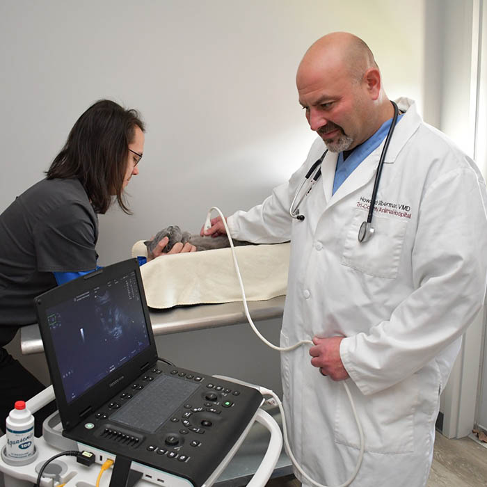
Recognizing Heart Ultrasound: Just How It Functions
A significant number of veterinary techniques currently make use of heart ultrasound as a crucial analysis tool for examining heart health and wellness in animals. This non-invasive strategy uses high-frequency sound waves to develop pictures of the heart's structure and function. During the treatment, a veterinary professional applies a gel to the pet's upper body and utilizes a transducer to produce ultrasound waves. These waves bounce off the heart and surrounding frameworks, creating real-time pictures on a monitor.Veterinarians can evaluate different aspects of heart health and wellness, consisting of chamber dimension, wall surface activity, and valve feature. Furthermore, cardiac ultrasound permits the detection of problems such as liquid buildup and congenital heart flaws. This strategy is vital for diagnosing problems that might not be noticeable through typical radiographs. By offering thorough info concerning the heart's anatomy and performance, cardiac ultrasound help in formulating effective treatment plans for pets experiencing cardiovascular disease.
The Value of CT Scans in Diagnosing Heart Conditions
Just how do CT scans enhance the diagnosis of heart conditions in vet medication? CT scans provide thorough cross-sectional pictures of the heart and surrounding structures, allowing veterinarians to envision intricate physiological partnerships. This imaging strategy is especially helpful in recognizing congenital heart defects, heart lumps, and irregularities in blood vessels. By making use of sophisticated imaging algorithms, CT scans can examine heart chamber dimensions and function, supplying a detailed view click reference that may be hard company website to achieve with standard methods.Additionally, CT angiography can envision blood flow and determine areas of stenosis or blockage, which is important for intending prospective treatments. The speed and accuracy of CT scans additionally facilitate quick diagnoses, essential in emergency situation situations. Eventually, the unification of CT scans into vet cardiology substantially enhances the precision of medical diagnoses, making it possible for targeted therapy plans and improving individual outcomes for animals experiencing heart conditions.
Comparing Ultrasound and CT Scan Strategies
While both ultrasound and CT scans are important devices in veterinary cardiology, they use unique advantages and restrictions that affect their usage in detecting heart disease. Ultrasound, or echocardiography, provides real-time imaging of the heart's framework and feature, permitting vets to assess heart chambers, shutoffs, and blood flow. It is especially efficient for examining conditions like congestive heart failing and cardiomyopathy. Ultrasound might be limited in visualizing certain anatomical structures due to individual size or obesity.In contrast, CT checks offer detailed cross-sectional photos of the heart and surrounding tissues, making them ideal for recognizing architectural problems, tumors, or vascular concerns. Although CT scans supply complete insights, they need sedation and might involve radiation direct exposure. Ultimately, the choice in between ultrasound and CT checks depends upon the details medical scenario, the patient's condition, and the info required for an exact medical diagnosis.
Therapy Options Available With Vet Cardiology
Vet cardiology uses an array of treatment options customized to resolve numerous heart disease in pets. Therapy plans commonly start with way of living modifications, consisting of diet adjustments and exercise changes, targeted at boosting overall heart wellness. Drugs play an essential function, with cardiologists prescribing medications such as diuretics, beta-blockers, and ACE inhibitors to enhance and manage signs and symptoms heart function.In a lot more extreme instances, interventional procedures, such as balloon valvuloplasty or stent positioning, might be needed to reduce obstructions or improve blood flow. For particular congenital heart problems, medical click here to read alternatives may be checked out to deal with structural issues. Furthermore, recurring monitoring and follow-up care are vital parts of a thorough therapy plan, permitting timely modifications based upon the family pet's response to treatment. Overall, vet cardiology focuses on offering effective, customized care to optimize the health and wellness and well-being of animal patients with heart disease.
Just how to Prepare Your Animal for a Cardiac Analysis
Preparing a family pet for a heart evaluation is vital to assure exact results and a smooth process. Owners need to first schedule the visit with the vet cardiologist and talk about any type of certain demands or issues. It is a good idea to keep food for at the very least 12 hours prior to the examination, as this helps improve imaging high quality throughout procedures like ultrasound or CT scans.Additionally, preserving a calm setting on the day of the consultation can help in reducing the pet's stress and anxiety. It is useful to bring along any pertinent clinical records, consisting of previous tests and medicines (CT Scans For Animals). Owners ought to also ensure that their pet is comfy and leashed throughout transport to the facility. Finally, acquainting themselves with the examination process can aid and ease concerns in asking informed inquiries throughout the assessment. By following these steps, owners can add considerably to the effectiveness of the heart examination
Frequently Asked Inquiries
The length of time Does a Cardiac Ultrasound or CT Check Take?
The duration of a heart ultrasound normally ranges from 30 to 60 mins, while a CT scan may take about 15 to half an hour. Variables such as the individual's problem can influence these time price quotes.

Exist Any Risks Connected With These Analysis Treatments?
Can I Remain With My Animal During the Procedure?
The veterinary facility's policy generally dictates whether animal owners can remain during treatments. While some facilities urge proprietor existence for comfort, others may require separation to assure safety and ideal problems for analysis imaging.
Just how much Do These Diagnostic Tests Generally Cost?
The costs of analysis examinations, such as ultrasound and CT scans, normally vary based on location and center. Commonly, rates range from a few hundred to over a thousand dollars, mirroring the intricacy and innovation involved.
What Is the Recuperation Refine After a Cardiac Evaluation?
The recuperation procedure after a cardiac evaluation involves monitoring the pet for any kind of instant reactions, making certain convenience, and limiting physical task. Veterinarians generally provide post-evaluation guidelines to assist animal owners during this essential recuperation period. Heart murmurs, numerous kinds of cardiomyopathy, and hereditary heart issues are amongst the most prevalent problems that veterinarians run into. A heart murmur is an abnormal noise produced by stormy blood flow within the heart. Cardiomyopathy incorporates a team of diseases impacting the heart muscle, leading to jeopardized cardiac feature in pets. Genetic heart defects stand for a significant classification of heart problems in pet dogs, distinct from obtained problems such as cardiomyopathy. Ultrasound, or echocardiography, provides real-time imaging of the heart's structure and feature, permitting veterinarians to examine heart chambers, shutoffs, and blood flow.